Home
/ Cross Section Of A Long Bone, Bone Cross Section High Res Stock Images Shutterstock, They primarily serve to protect tendons.
Cross Section Of A Long Bone, Bone Cross Section High Res Stock Images Shutterstock, They primarily serve to protect tendons.
Cross Section Of A Long Bone, Bone Cross Section High Res Stock Images Shutterstock, They primarily serve to protect tendons.. Bone test anatomy and physiology 12 photos of the bone test anatomy and physiology anatomy and physiology bone lab test, anatomy and physiology bone markings test, anatomy and physiology bone practical test, anatomy and physiology bone tissue test, anatomy and physiology test on bone tissue, bone, anatomy and. Compact bone is the denser, stronger of the two types of bone tissue ( (figure) ). Shop the edit of floral dresses, dream jeans and fresh shoes now, and stay tuned for a lot more exciting topshop stuff to come. 320 × 160 pixels | 640 × 320 pixels | 1,024 × 512 pixels | 1,280 × 640 pixels | 1,000 × 500 pixels. Would it be a good thing to show the epiphyseal plate?
Bone markings the surface features of bones vary considerably, depending on the function and location in the body. We feature 65,400,000 royalty free photos, 337,000 stock footage clips, digital videos, vector clip art images, clipart pictures, background graphics, medical illustrations. The central tubular region of the bone, called the diaphysis, flares outward near the end to form the metaphysis, which contains a largely cancellous, or spongy, interior. An outer 'fibrous layer' containing mainly fibroblasts, and an inner 'cambium layer' containing progenitor cells. Internal structure of a human long bone, with a magnified cross section of the interior.
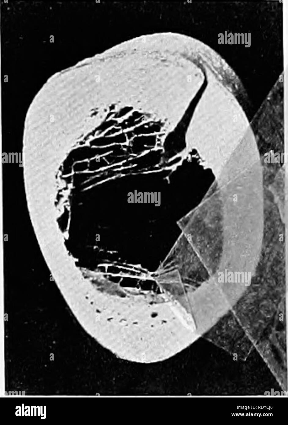
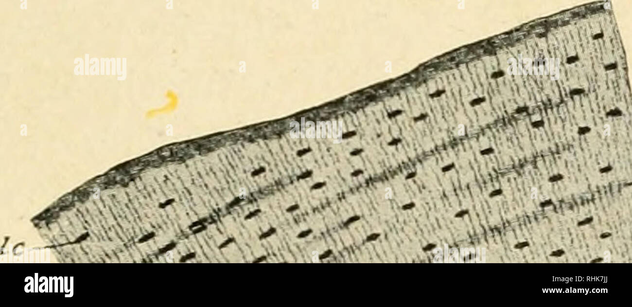
The Anatomy Of The Domestic Animals Veterinary Anatomy Fig 2 Cross Section Of Proximal Third Of Shaft Of Right Humerus Of Horse Fig 3 Cross Section Of Distal Third Of Shaft Of from c8.alamy.com Eliminate sudden changes of direction and influx of one stream into another. In long bones, as you move from the outer cortical compact bone to the inner medullary cavity, the bone transitions to spongy bone. If you look at the cross section of a long bone under a microscope, the rings of bone immediately internal to the periosteum of the bone are called external circumferential lamellae. Muscle attachments are visible along the outer surface. The section 8 housing program helps nee. The compact bone is made up of osteon. Bone in arm pictures 12 photos of the bone in arm pictures bone cancer arm pictures, pictures of bone cancer in arm, bone, bone cancer arm pictures, pictures of bone cancer in arm. There are trabeculae in spongy bone which gives its sponge like appearance.
Examples of flat bones include ribs, scapulae and skull bones.
There are trabeculae in spongy bone which gives its sponge like appearance. Bone markings the surface features of bones vary considerably, depending on the function and location in the body. Internal structure of a human long bone, with a magnified cross section of the interior. We feature 65,400,000 royalty free photos, 337,000 stock footage clips, digital videos, vector clip art images, clipart pictures, background graphics, medical illustrations. Spongy bone, also known as cancellous bone or trabecular bone, looks like a sponge under the microscope. The central tubular region of the bone, called the diaphysis, flares outward near the end to form the metaphysis, which contains a largely cancellous, or spongy, interior. Size of this png preview of this svg file: Body size standardization was done, using the following equations: Transverse cross section of compact bone tissue; Why is the marrow red? They primarily serve to protect tendons. And why does the marrow stop where it does, and so sharply? Would it be a good thing to show the epiphyseal plate?
Spongy bone, also known as cancellous bone or trabecular bone, looks like a sponge under the microscope. An approach to model the effect of exercise on the growth of mammal long bones is described. End of a long bone. In addition, cortical bone thickness at anterior, posterior, medial, and lateral parts of the bone section was measured. An outer 'fibrous layer' containing mainly fibroblasts, and an inner 'cambium layer' containing progenitor cells.
Pediatric Skeletal Growth from www.fairview.org Section 8 refers to the section 8 housing program, also called the housing choice voucher program. Eliminate sudden changes of direction and influx of one stream into another. The central tubular region of the bone, called the diaphysis, flares outward near the end to form the metaphysis, which contains a largely cancellous, or spongy, interior. If you look at the cross section of a long bone under a microscope, the rings of bone immediately internal to the periosteum of the bone are called external circumferential lamellae. Related posts of cross section of a long bone bone test anatomy and physiology. Body size standardization was done, using the following equations: It consists of two layers; Terms in this set (3) epiphysis.
Body size standardization was done, using the following equations:
Body size standardization was done, using the following equations: Terms in this set (3) epiphysis. Marrow in the shaft of long bones is typically yellow, with red marrow in the head through the cancellous bone. Chapter 6 bones and skeletal tissues flashcards quizlet. An approach to model the effect of exercise on the growth of mammal long bones is described. If you look at the cross section of a long bone under a microscope, the rings of bone immediately internal to the periosteum of the bone are called external circumferential lamellae. They primarily serve to protect tendons. End of a long bone. It can be found under the periosteum and in the diaphyses of long bones, where it provides support and protection. This is a file from the wikimedia commons. This is known as the periosteum. I don't find it enhances the image. There are trabeculae in spongy bone which gives its sponge like appearance.
Internal structure of a human long bone, with a magnified cross section of the interior. Compact bone is the denser, stronger of the two types of bone tissue ( (figure) ). In addition, cortical bone thickness at anterior, posterior, medial, and lateral parts of the bone section was measured. Marrow in the shaft of long bones is typically yellow, with red marrow in the head through the cancellous bone. The section 8 housing program helps nee.
The Biology Of The Frog Frogs Histology Of The Frog 127 Im Compact Bone Which Is Very Firm And Dense And Spongy Or Cancellous Bone Which Is Made Up Of from c8.alamy.com Spongy bone also contains osteocytes housed in lacunae, but they are not arranged in concentric circles. In addition, cortical bone thickness at anterior, posterior, medial, and lateral parts of the bone section was measured. Pdb05005 fotosearch stock photography and stock footage helps you find the perfect photo or footage, fast! Cartilaginous area at the ends of long bones where lengthwise growth takes place in the immature skeleton. Section 8 refers to the section 8 housing program, also called the housing choice voucher program. The structure of a long bone allows for the best visualization of all of the parts of a bone (figure 1). Two types of bone tissues in cross section of a long bone : In long bones, as you move from the outer cortical compact bone to the inner medullary cavity, the bone transitions to spongy bone.
Spongy bone also contains osteocytes housed in lacunae, but they are not arranged in concentric circles.
We feature 65,400,000 royalty free photos, 337,000 stock footage clips, digital videos, vector clip art images, clipart pictures, background graphics, medical illustrations. Spongy bone, also known as cancellous bone or trabecular bone, looks like a sponge under the microscope. A long bone has two parts: Why is the marrow red? Long bones are longer than they are wide and are the major bones of the limbs. Examples of flat bones include ribs, scapulae and skull bones. In long bones, as you move from the outer cortical compact bone to the inner medullary cavity, the bone transitions to spongy bone. The structure of a long bone allows for the best visualization of all of the parts of a bone (figure 1). 320 × 160 pixels | 640 × 320 pixels | 1,024 × 512 pixels | 1,280 × 640 pixels | 1,000 × 500 pixels. Transverse cross section of compact bone tissue; It can be found under the periosteum and in the diaphyses of long bones, where it provides support and protection. An outer 'fibrous layer' containing mainly fibroblasts, and an inner 'cambium layer' containing progenitor cells. (area/long bone length 3) ∗ 10 8.
An approach to model the effect of exercise on the growth of mammal long bones is described cross section of a bone. Spongy bone, also known as cancellous bone or trabecular bone, looks like a sponge under the microscope.